

Pelvic floor ultrasound in Mérida
A safe, radiation-free scan that shows in real time how the bladder, uterus, rectum and the muscles that support them are working. It uses 3D and 4D imaging to diagnose incontinence, prolapse or pelvic pain and to help plan treatment.
Performed by: Dr. David Peba Ceballos, radiologist certified by the Mexican Council of Radiology and Imaging (specialty license no. 12855636)
Duration: 30 to 45 minutes
Preparation: general hygiene only
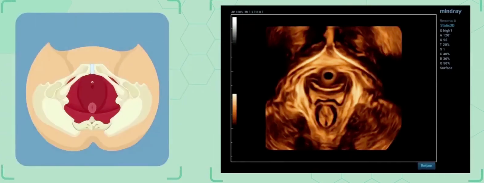
The goal of this assessment is to obtain an anatomical and functional map of the pelvic floor, to give each patient better guidance and to plan rehabilitation or surgery more precisely.
Frequently asked questions
What does it assess?
- Dysfunction: urinary incontinence (stress or urge) and fecal incontinence.
- Pelvic organ prolapse: descent of the bladder (cystocele), uterus, vagina or rectum (rectocele).
- Muscle function: how the levator ani muscles and the sphincter contract and relax.
- Organ position: bladder, urethra and rectum, and how they move during urination or defecation.
- Support structures: ligaments and fascia.
- Surgical meshes: to check their position and function after surgery.
What are the advantages of this ultrasound?
- Dynamic and real time: shows how the pelvic floor works, not just its shape.
- Safe and non-invasive: no radiation.
- Versatile: complements other tests and helps guide rehabilitation.
- Simple preparation: no fasting is needed, and results are quick.
Who is it for?
- Women and men with incontinence, prolapse, pelvic pain or sexual dysfunction.
- Patients in pelvic floor rehabilitation, after childbirth or after surgery.
Book your pelvic floor ultrasound
The scan is performed personally by Dr. David Peba Ceballos, a radiologist certified by the Mexican Council of Radiology and Imaging. Message us on WhatsApp and we will send you the preparation instructions, availability and price.